Services on Demand
Journal
Article
Indicators
Related links
-
 Cited by Google
 -
 Similars in Google
Share
SAMJ: South African Medical Journal
On-line version ISSN 2078-5135Print version ISSN 0256-9574
SAMJ, S. Afr. med. j. vol.99 n.12 Pretoria Dec. 2009
SAMJ FORUM
CLINICAL IMAGES
Retained glass fragments in body tissues
D J Emby*
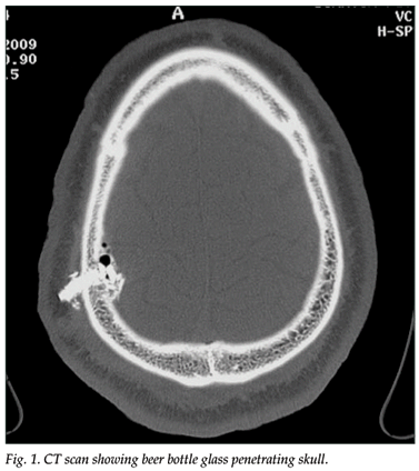
A 34-year-old man presented with a fragment of glass protruding from his scalp after having been struck with a beer bottle that had broken on impact. The glass was visible on plain film radiographs, but its degree of penetration could not be determined. A computed tomography (CT) scan showed a fragment of glass penetrating the calvarium, glass shards in the scalp and intracranially, and tiny intracranial bone fragments and small air loculations (Fig. 1). A flap containing the embedded glass fragment was removed and the wound debrided.

The medical literature frequently fails to distinguish between different types of glass as a foreign body, and often states that all glass is visible on X-rays.1 However, small shards of glass may not be visible on plain film radiographs because the radio-densities of different types of glass differ qualitatively.2 Beer bottle glass is usually visible on plain radiographs, but is much less dense than on a CT scan (Fig. 2). Shattered windshield glass is clearly visible on plain radiographs, as its manufacture renders it significantly radio-opaque (Fig. 3). Windowpane glass appears to be the least radio-opaque and can be difficult to visualise in the soft tissues on plain radiographs.


To demonstrate the advantage of CT over plain film X-rays, a saline bag simulating soft tissue was placed over a sample of windowpane glass and a plain film radiograph was obtained (Fig. 4). This glass, which is faintly visible on either side of the bag, merges in density with the saline and can only be differentiated from the bag at its lateral edges. On a CT scan the thin layer of glass is clearly visible underneath the saline bag (Fig. 5), illustrating that the CT scan is much more sensitive than plain films in differentiating ranges of density. CT also has superior spatial resolution within soft tissue, and within bone (as shown in Fig. 1). CT is therefore the preferred imaging modality for detecting shards of glass in the soft tissues when these are not clearly visible or inadequately anatomically defined on plain film radiographs.


The greater sensitivity of CT is useful in clinical conditions that previously presented diagnostic dilemmas, e.g. ureteric calculi of insufficient density to be visible on plain films and impacted fish bones in the hypopharynx and cervical oesophagus, which may not be visible on plain films (Fig. 6).3

1. Rothschild MA, Karger B, Schneider V. Puncture wounds caused by glass mistaken for with stab wounds with a knife. Forensic Sci Int 2001; 121: 161-165.
2. Andrew WK. Sonographic diagnosis of foreign bodies of the distal extremities. AJR Am J Roentgenol 1987; 149: 864.
3. Watanabe K, Kikuchi T, Katori Y, et al. The usefulness of computed tomography in the diagnosis of impacted fish bones in the oesophagus. J Laryngol Otol 1998; 112(4): 360-364.
Dr Emby graduated from the University of the Witwatersrand and trained in radiology on the Durban circuit. He subsequently became Consultant Radiologist to Rand Mutual Hospital and the Chamber of Mines and is currently Consultant Radiologist to AngloGold Ashanti Health. Special interests include trauma, industrial chest disease, and optimal use of imaging modalities in order to contain radiation exposure in medical imaging.
* Corresponding author: D J Emby (demby@anglogoldashanti.com)


