



Services on Demand
Journal

Article
 English (pdf)
 Article in xml format
 Article references

 Automatic translation
 Send this article by e-mail
Indicators
Related links
-
 Cited by Google
-
 Similars in Google
Share

 Permalink

SA Journal of Radiology
On-line version ISSN 2078-6778Print version ISSN 1027-202X
S. Afr. J. radiol. (Online) vol.29 n.1 Johannesburg 2025
https://doi.org/10.4102/sajr.v29i1.3246
CASE SERIES
Imaging Klippel-Trenaunay and Parkes Weber syndromes: A unilateral limb overgrowth case series
Himanshu Nirwal; Priya Singh; Surya P. Singh; Anit Parihar
Department of Radiodiagnosis, King George's Medical University, Lucknow, India
ABSTRACT
This case series highlights the importance of multimodal imaging in distinguishing Klippel-Trenaunay syndrome (KTS) from Parkes Weber syndrome (PWS) and guiding management. Both KTS and PWS are rare congenital vascular disorders characterised by the classic triad of cutaneous vascular malformations, venous varicosities and limb overgrowth. The three cases in this report highlight the spectrum of vascular malformations and their imaging characteristics, emphasising the importance of flow assessment in distinguishing low-flow venous malformations (KTS) from high-flow arteriovenous malformations (PWS). Multimodal cross-sectional imaging is essential for accurate diagnosis and treatment planning in vascular overgrowth syndromes.
CONTRIBUTION: This case series illustrates the critical role of multimodal imaging in differentiating Klippel-Trenaunay syndrome from Parkes Weber syndrome. It emphasizes the distinct imaging findings, particularly vascular flow characteristics, which are essential for accurate diagnosis, guiding appropriate therapeutic interventions, and preventing serious complications
Keywords: Klippel-Trenaunay syndrome; Parkes Weber syndrome; vascular malformations; multimodal imaging; unilateral lower limb enlargement.
Introduction
Klippel-Trenaunay syndrome (KTS) represents a rare congenital disorder within the PIK3CA-related overgrowth spectrum (PROS), while Parkes Weber syndrome (PWS) occurs because of a RASA1 gene mutation.1,2 Both are characterised by the classic triad of cutaneous vascular malformations, venous varicosities and limb overgrowth. The estimated incidence ranges from 3 to 5 per 100 000 individuals, with unilateral lower limb involvement occurring in approximately 95% of cases.3
The fundamental distinction between these syndromes lies in the vascular flow characteristics: KTS presents with low-flow venous and lymphatic malformations, while PWS involves high-flow arteriovenous malformations with direct arteriovenous communications.4 This differentiation is crucial as PWS carries significantly higher risks of complications including congestive heart failure, bleeding and thromboembolic events.5 Other congenital disorders that can cause limb length discrepancy with vascular malformations include CLOVES syndrome, Proteus syndrome and fibroadipose vascular anomaly (FAVA). They can be differentiated by their different phenotypic features and further confirmed by genetic testing.4
Recent advances in cross-sectional imaging, particularly contrast-enhanced CT angiography (CTA) and MRA, have revolutionised the diagnostic approach to these complex vascular disorders.6 These modalities provide detailed anatomical information, flow characteristics assessment, and comprehensive evaluation of deep venous anatomy, which is essential for surgical planning and patient management.7
The management of both syndromes remains largely conservative, focusing on symptomatic relief and complication prevention. However, accurate diagnosis is paramount as therapeutic interventions differ significantly between the two conditions.8 While KTS patients may benefit from sclerotherapy and surgical debulking, PWS often requires embolisation procedures because of the high-flow nature of the vascular malformations.9 This case series aims to illustrate the imaging spectrum of KTS and PWS, emphasising the critical role of multimodal imaging in accurate diagnosis and therapeutic planning.
Ethical considerations
Written informed consent was obtained from the patients.
Case presentations
Case 1: Parkes Weber syndrome
A 15-year-old female presented with progressive hypertrophy of the left lower limb and bluish discolouration of the foot region, present since birth. Clinical examination revealed limb overgrowth with multiple tortuous vessels throughout the entire limb, producing palpable thrills indicative of high-flow vascular malformations.
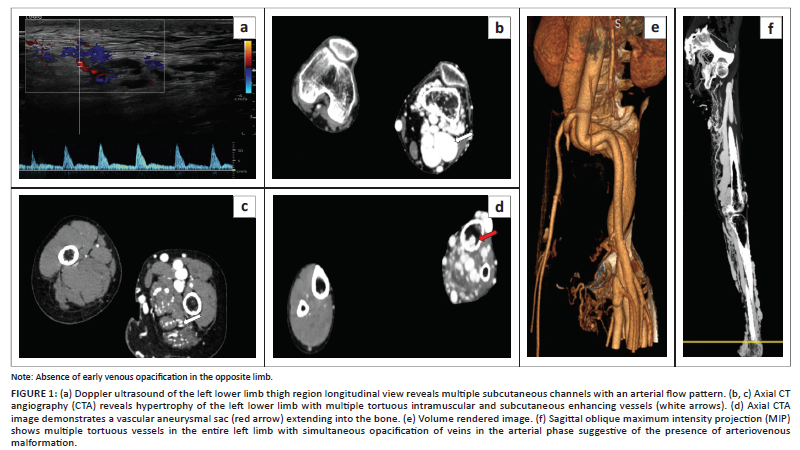
Plain radiography demonstrated diffuse osteopenia of the left lower limb bones without structural deformities. Colour Doppler ultrasonography (Figure 1a) revealed multiple tortuous vessels with both venous and arterialised flow patterns, suggesting high-flow arteriovenous malformations.
Computed tomography angiography of the lower limb (non-contrast followed by arterial and venous phase angiography) (Figure 1 b-f) confirmed extensive arteriovenous malformations with multiple tortuous vessels in the subcutaneous and intramuscular regions. The pathognomonic finding was early venous filling during the arterial phase of contrast enhancement, confirming arteriovenous shunting. Additional findings included diffuse cortical thinning of the left lower limb bones with focal erosions and intraosseous extension of the vascular malformations.
The combination of high-flow arteriovenous malformations with early venous filling distinguished this case as PWS. The patient underwent successful transcatheter embolisation therapy with n-Butyl-2-cyanoacrylate (nBCA) adhesive in staged procedures, followed by surgical debulking.
Case 2: Klippel-Trenaunay syndrome
A 20-year-old male presented with left lower limb swelling, hypertrophy and patchy skin discolouration since childhood (Figure 2a). Physical examination revealed prominent superficial varicosities and non-tender, compressible soft tissue masses without ulcerations or bone deformities.
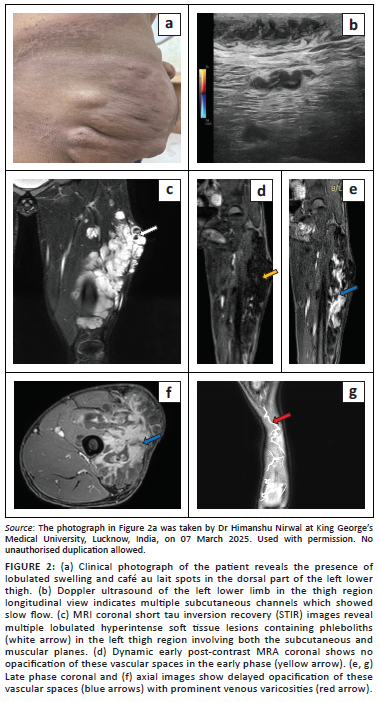
Colour Doppler ultrasonography (Figure 2b) demonstrated extensive lobulated vascular spaces in the superficial and deep muscular planes with characteristic slow venous flow. Multiple venous varicosities were identified along the lateral aspect of the limb, consistent with low-flow venous malformations. The deep veins were patent.
Lower limb MRI (T1 W axial, T2 W axial, T1W fat saturated axial, short tau inversion recovery [STIR] coronal and axial time-resolved imaging of contrast kinetics [TRICKS] angiography) (Figure 2 c-g) revealed multiple clusters of T2-hyperintense vascular spaces showing delayed contrast filling typical of venous malformations. Subcutaneous hypertrophy of the left lower limb was evident. The absence of early opacification of the vascular channels and the presence of delayed venous enhancement confirmed the low-flow nature of the malformations.
All three components of the KTS triad were present, establishing the diagnosis. Further confirmation was achieved through genetic testing. The patient was successfully managed with multiple sessions of ultrasound-guided foam sclerotherapy with sodium tetradecyl sulfate and remains on regular follow-up.
Case 3: Klippel-Trenaunay syndrome with complications
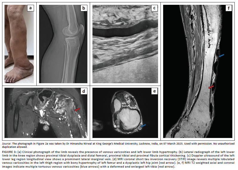
A 16-year-old male presented with progressive left limb enlargement, deformity, and multiple cutaneous port-wine stains. Physical examination revealed prominent superficial varicosities on both the medial and lateral aspects of the lower limb (Figure 3a). The limb showed reduced mobility at the hip, knee and ankle joints with tibial and fibular bowing. Small ulcers were present in the foot.
Plain radiography demonstrated bowing deformities of the tibia and fibula with diffuse bone thickening and soft tissue overgrowth (Figure 3b). The articular surface of the left hip joint appeared flattened and dysplastic, with ankylosis of the distal tibia and fibula.
Colour Doppler ultrasonography (Figure 3c) revealed multiple superficial venous channels and varicosities, including a prominent venous channel along the lateral thigh consistent with a lateral marginal vein. The deep veins were patent. MRI (T1 W axial, T2 W axial, T1W fat saturated axial, STIR coronal and axial, high resolution fat supressed time resolved MRA, venography) (Figure 3d-f) confirmed multiple tortuous venous varicosities with a deformed and hypertrophied left tibia and fibula. The left femur demonstrated diffuse thickening with dysplastic hip joint changes. Subcutaneous hypertrophy was prominent throughout the affected limb.
The imaging findings confirmed all three components of the KTS triad with significant skeletal complications. Management included multiple sessions of ultrasound-guided foam sclerotherapy with sodium tetradecyl sulfate and continued orthopaedic follow-up for skeletal deformities.
Discussion
This case series demonstrates the critical importance of a comprehensive imaging evaluation in differentiating KTS from PWS and guiding appropriate therapeutic management. The fundamental distinction between these syndromes lies in vascular flow characteristics, which directly impact treatment strategies and patient outcomes.10
Imaging characteristics and diagnostic criteria
The hallmark imaging feature distinguishing PWS from KTS is the presence of high-flow arteriovenous malformations with early venous filling during the arterial phase of contrast enhancement.11 This was clearly demonstrated in case 1, where CTA revealed rapid arteriovenous shunting with early venous opacification. In contrast, cases 2 and 3 showed the characteristic delayed venous filling pattern, typical of low-flow venous malformations in KTS.12
Recent literature emphasises that KTS occurs as a result of somatic mutations in the PIK3CA gene, forming part of the PROS disorders.2 The diagnostic criteria have evolved from the classical triad to a more comprehensive understanding that includes the assessment of vascular flow characteristics, which the presented cases clearly illustrate.13
Parkes Weber syndrome remains less well-characterised in radiological literature, necessitating broader recognition and documentation.14,15 Recent genetic studies have identified RASA1 mutations as the primary cause of PWS, distinguishing it from the PIK3CA mutations associated with KTS, and this genetic differentiation correlates with distinct imaging phenotypes that warrant systematic documentation.16,17 The PWS typically presents with more aggressive vascular malformations, higher rates of cardiac complications because of high-flow shunting and increased bleeding risk compared to KTS, making early and accurate radiological diagnosis crucial for patient outcomes.18,19 Furthermore, the radiological assessment of the treatment response in PWS requires specialised protocols, as conventional response criteria may not adequately capture the complex haemodynamic changes following embolisation.20
Multimodal imaging approach
The cases demonstrate the value of a structured imaging protocol combining multiple modalities. Colour Doppler ultrasonography serves as the initial screening tool, providing real-time flow assessment and distinguishing between high-flow and low-flow malformations.21 The patency of deep veins can be confirmed on ultrasound, which is important for treatment planning. However, cross-sectional imaging with CTA or MRA is essential for complete anatomical mapping and surgical planning.22 Moreover, radiologists can direct the type of genetic testing based on imaging findings for confirmation. Digital subtraction angiography can be performed where therapeutic intervention is planned.
The Egyptian study by Kaddah et al. proposed a comprehensive radiological protocol involving CT scanograms, MRI, MRA and magnetic resonance venography as routine studies, which aligns with the approaches in the cases presented.7 Their series of 20 patients demonstrated that this protocol significantly aids in treatment planning and long-term follow-up.
Skeletal and soft tissue involvement
All three cases in this series demonstrated varying degrees of limb overgrowth, with case 3 showing the most severe skeletal complications including bowing deformities and joint ankylosis. The radiological assessment of skeletal involvement is crucial for orthopaedic planning and long-term management.23
The presence of intraosseous extension of vascular malformations, as seen in case 1, represents a more severe form of the disease and may contribute to bone weakening and deformities.24 This finding has been reported in the literature as an indicator of disease severity and potential complications.
Role of radiology in management and treatment
Beyond diagnostic differentiation, radiology plays a pivotal role in the multidisciplinary management and treatment of both KTS and PWS. In PWS, interventional radiology serves as the primary therapeutic modality, with transcatheter embolisation using various embolic agents including nBCA, coils and plugs being the treatment of choice for high-flow arteriovenous malformations.25,26,27 Pre-procedural angiographic mapping is essential for identifying feeding arteries, nidus characteristics, and venous drainage patterns, which directly influence embolisation strategy and outcomes.28
In KTS, ultrasound-guided percutaneous sclerotherapy represents the mainstay of interventional treatment, with foam sclerotherapy using sodium tetradecyl sulfate or polidocanol, being particularly effective for superficial venous malformations.21,29 In addition, endovenous laser ablation and radiofrequency ablation may be employed for incompetent superficial veins, while balloon-occluded retrograde transvenous obliteration can be utilised for complex deep venous malformations.30,31 Cross-sectional imaging guidance ensures precise targeting of lesions while avoiding critical structures, and post-treatment imaging surveillance is crucial for assessing therapeutic response and detecting complications such as skin necrosis or deep vein thrombosis.32
Conclusion
This case series demonstrates that multimodal cross-sectional imaging is indispensable for accurate diagnosis and treatment planning in KTS and PWS. The critical differentiating feature between these conditions is the vascular flow characteristics, with PWS showing high-flow arteriovenous malformations and early venous filling, while KTS demonstrates low-flow venous malformations with delayed contrast enhancement. Early recognition and appropriate imaging evaluation of these rare, but complex vascular disorders, ensure optimal patient outcomes and prevent serious complications.
The cases illustrate the spectrum of imaging findings from mild venous malformations to severe complications involving skeletal deformities and intraosseous extension. The comprehensive imaging approach combining Doppler ultrasonography, CTA and MRI provides essential information for therapeutic decision-making and long-term patient management. Future studies with larger cohorts exploring standardised imaging protocols and advanced imaging techniques would be beneficial for establishing evidence-based diagnostic criteria.
Acknowledgements
Competing interests
The authors declare that they have no personal or financial relationship that may have inappropriately influenced the writing of this article.
Authors' contributions
H.N. and P.S. were the primary authors and have contributed equally, sharing first authorship. S.P.S. and A.P. supervised and contributed equally to the final version of the article.
Funding information
This research received no specific grant from any funding agency in the public, commercial or not-for-profit sectors.
Data availability
Data sharing is available from the corresponding author, P.S., upon request.
Disclaimer
The views and opinions expressed in this article are those of the authors and are the product of professional research. The article does not necessarily reflect the official policy or position of any affiliated institution, funder, agency, or that of the publisher. The authors are responsible for this article's results, findings, and content.
References
1. Turner VL, Kearns C, Wattamwar K, McKenney AS. Klippel-Trenaunay syndrome. RadioGraphics. 2022;42(6):E167-E168. https://doi.org/10.1148/rg.220150 [ Links ]
2. Vahidnezhad H, Youssefian L, Uitto J. Klippel-Trenaunay syndrome belongs to the PIK3CA-related overgrowth spectrum (PROS). Exp Dermatol. 2016;25(1):17-19. https://doi.org/10.1111/exd.12826 [ Links ]
3. Das R, Kumar I, Verma A, Shukla RC. Spectrum of imaging findings in Klippel-Trenaunay syndrome affecting lower limbs: A report of three cases. Egypt J Radiol Nucl Med. 2019;50:104. https://doi.org/10.1186/s43055-019-0123-7 [ Links ]
4. Bertino F, Braithwaite KA, Hawkins CM, et al. Congenital limb overgrowth syndromes associated with vascular anomalies. RadioGraphics. 2019;39(2):491-515. https://doi.org/10.1148/rg.2019180136 [ Links ]
5. International Society for the Study of Vascular Anomalies (ISSVA). Classification for vascular anomalies [homepage on the Internet]. [cited 2022 Jun 15]. Available from: https://www.issva.org/UserFiles/file/ISSVA-Classification-2018.pdf [ Links ]
6. Nozaki T, Nosaka S, Miyazaki O, et al. Syndromes associated with vascular tumors and malformations: A pictorial review. RadioGraphics. 2013;33(1):175-195. https://doi.org/10.1148/rg.331125052 [ Links ]
7. Kaddah RO, Lotfi U, Haggag M, Abd EL, Ghani H. The role of radiology in the planning management of Klippel Trenaunay Syndrome (KTS). Egypt J Radiol Nucl Med. 2011;42:201-210. https://doi.org/10.1016/j.ejrnm.2011.06.002 [ Links ]
8. John PR. Klippel-Trenaunay syndrome. Tech Vasc Interv Radiol. 2019;22(4):100634. https://doi.org/10.1016/j.tvir.2019.100634 [ Links ]
9. Luks VL, Kamitaki N, Vivero MP, et al. Lymphatic and other vascular malformative/overgrowth disorders are caused by somatic mutations in PIK3CA. J Pediatr. 2015;166(4):1048-1054. https://doi.org/10.1016/j.jpeds.2014.12.069 [ Links ]
10. Oduber CE, Van der Horst CM, Hennekam RC. Klippel-Trenaunay syndrome: Diagnostic criteria and hypothesis on etiology. Ann Plast Surg. 2008;60:217-222. https://doi.org/10.1097/SAP.0b013e318062abc1 [ Links ]
11. Jacob AG, Driscoll DJ, Shaughnessy WJ, Stanson AW, Clay RP, Gloviczki P. Klippel Trenaunay syndrome: Spectrum and management. Mayo Clin Proc. 1998;73:28-36. https://doi.org/10.1016/S0025-6196(11)63615-X [ Links ]
12. Cha SH, Romeo MA, Neutze JA. Visceral manifestations of Klippel-Trenaunay syndrome. RadioGraphics. 2005;25:1694-1697. https://doi.org/10.1148/rg.256055042 [ Links ]
13. Berry SA, Peterson C, Mize W, et al. Klippel-Trenaunay syndrome. Am J Med Genet. 1998;79:319-326. https://doi.org/10.1002/(SICI)1096-8628(19981002)79:4%3C319::AID-AJMG15%3E3.0.CO;2-U [ Links ]
14. Revencu N, Boon LM, Mulliken JB, et al. Parkes Weber syndrome, vein of Galen aneurysmal malformation, and other fast-flow vascular anomalies are caused by RASA1 mutations. Hum Mutat. 2008;29(7):959-965. https://doi.org/10.1002/humu.20746 [ Links ]
15. Burrows PE, Mitri RK, Alomari A, et al. Percutaneous sclerotherapy of lymphatic malformations with doxycycline. Lymphat Res Biol. 2008;6(3-4):209-216. https://doi.org/10.1089/lrb.2008.1004 [ Links ]
16. Amyere M, Revencu N, Helaers R, et al. Germline loss-of-function mutations in EPHB4 cause a second form of capillary malformation-arteriovenous malformation (CM-AVM2) deregulating RAS-MAPK signaling. Circulation. 2017;136(11):1011-1020. https://doi.org/10.1161/CIRCULATIONAHA.116.026886 [ Links ]
17. Soblet J, Limaye N, Uebelhoer M, et al. Variable somatic TIE2 mutations in half of sporadic venous malformations. Mol Syndromol. 2013;4(4):179-183. https://doi.org/10.1159/000348327 [ Links ]
18. Gloviczki P, Duncan A, Kalra M, et al. Vascular malformations: An update. Perspect Vasc Surg Endovasc Ther. 2009;21(2):133-148. https://doi.org/10.1177/1531003509343019 [ Links ]
19. Wouters V, Limaye N, Uebelhoer M, et al. Hereditary cutaneomucosal venous malformations are caused by TIE2 mutations with widely variable hypofunction effects. Eur J Hum Genet. 2010;18(4):414-420. https://doi.org/10.1038/ejhg.2009.193 [ Links ]
20. Markl M, Frydrychowicz A, Kozerke S, Hope M, Wieben O. 4D flow MRI. J Magn Reson Imaging. 2012;36(5):1015-1036. https://doi.org/10.1002/jmri.23632 [ Links ]
21. Ernemann U, Kramer U, Miller S, et al. Current concepts in the classification, diagnosis and treatment of vascular anomalies. Eur J Radiol. 2010;75(1):2-11. https://doi.org/10.1016/j.ejrad.2010.04.009 [ Links ]
22. Herborn CU, Goyen M, Lauenstein TC, et al. Comprehensive time-resolved MRA of peripheral vascular malformation. AJR Am J Roentgenol. 2003;181:729-735. https://doi.org/10.2214/ajr.181.3.1810729 [ Links ]
23. Delis KT, Gloviczki P, Wennberg PW, Rooke TW, Driscoll DJ. Hemodynamic impairment, venous segmental disease, and clinical severity scoring in limbs with Klippel-Trenaunay syndrome. J Vasc Surg. 2007;45(3):561-567. https://doi.org/10.1016/j.jvs.2006.11.032 [ Links ]
24. Guidera KJ, Brinker MR, Kousseff BG, et al. Overgrowth management in Klippel-Trenaunay-Weber and Proteus syndromes. J Pediatr Orthop. 1993;13:459-466. https://doi.org/10.1097/01241398-199307000-00009 [ Links ]
25. Yakes WF, Rossi P, Odink H. How I do it. Arteriovenous malformation management. Cardiovasc Intervent Radiol. 1996;19(2):65-71. https://doi.org/10.1007/BF02563895 [ Links ]
26. Jackson JE, Mansfield AO, Allison DJ. Treatment of high-flow vascular malformations by venous embolization aided by flow occlusion techniques. Cardiovasc Intervent Radiol. 1996;19(5):323-328. https://doi.org/10.1007/BF02570183 [ Links ]
27. Do YS, Yakes WF, Shin SW, et al. Ethanol embolization of arteriovenous malformations: Interim results. Radiology. 2005;235(2):674-682. https://doi.org/10.1148/radiol.2352040449 [ Links ]
28. Cabrera J, Cabrera J Jr, Garcia-Olmedo MA. Treatment of venous malformations with sclerosant in microfoam form. Arch Dermatol. 2003;139(11):1409-1416. https://doi.org/10.1001/archderm.139.11.1409 [ Links ]
29. Gloviczki P, Driscoll DJ. Klippel-Trenaunay syndrome: Current management. Phlebology. 2007;22(6):291-298. https://doi.org/10.1258/026835507782655209 [ Links ]
30. Mattassi R, Loose DA, Vaghi M. Hemangiomas and vascular malformations: An atlas of diagnosis and treatment. 2nd ed. Milan: Springer-Verlag; 2015. [ Links ]
31. Lee KB, Kim DI, Oh SK, et al. Incidence of soft tissue injury and neuropathy after foam sclerotherapy for venous malformations. J Vasc Surg. 2008;48(5):1286-1291. https://doi.org/10.1016/j.jvs.2008.06.058 [ Links ]
32. Wilson CL, Song LM, Chua H, et al. Bleeding from cavernous angiomatosis of the rectum in Klippel-Trenaunay syndrome: Report of three cases and literature review. Am J Gastroenterol. 2001;96:2783-2788. https://doi.org/10.1016/S0002-9270(01)02673-9 [ Links ]
 Correspondence:
Priya Singh
priyasingh@kgmcindia.edu
Received: 02 July 2025
Accepted: 16 Sept. 2025
Published: 19 Nov. 2025